
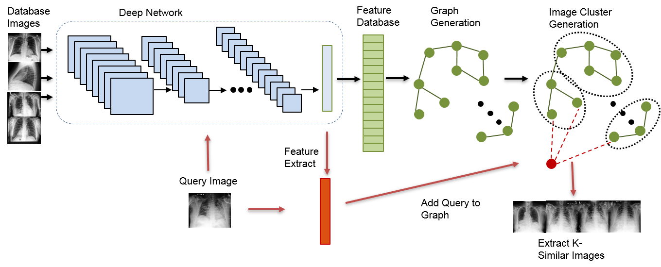
Large scale medical image retrieval
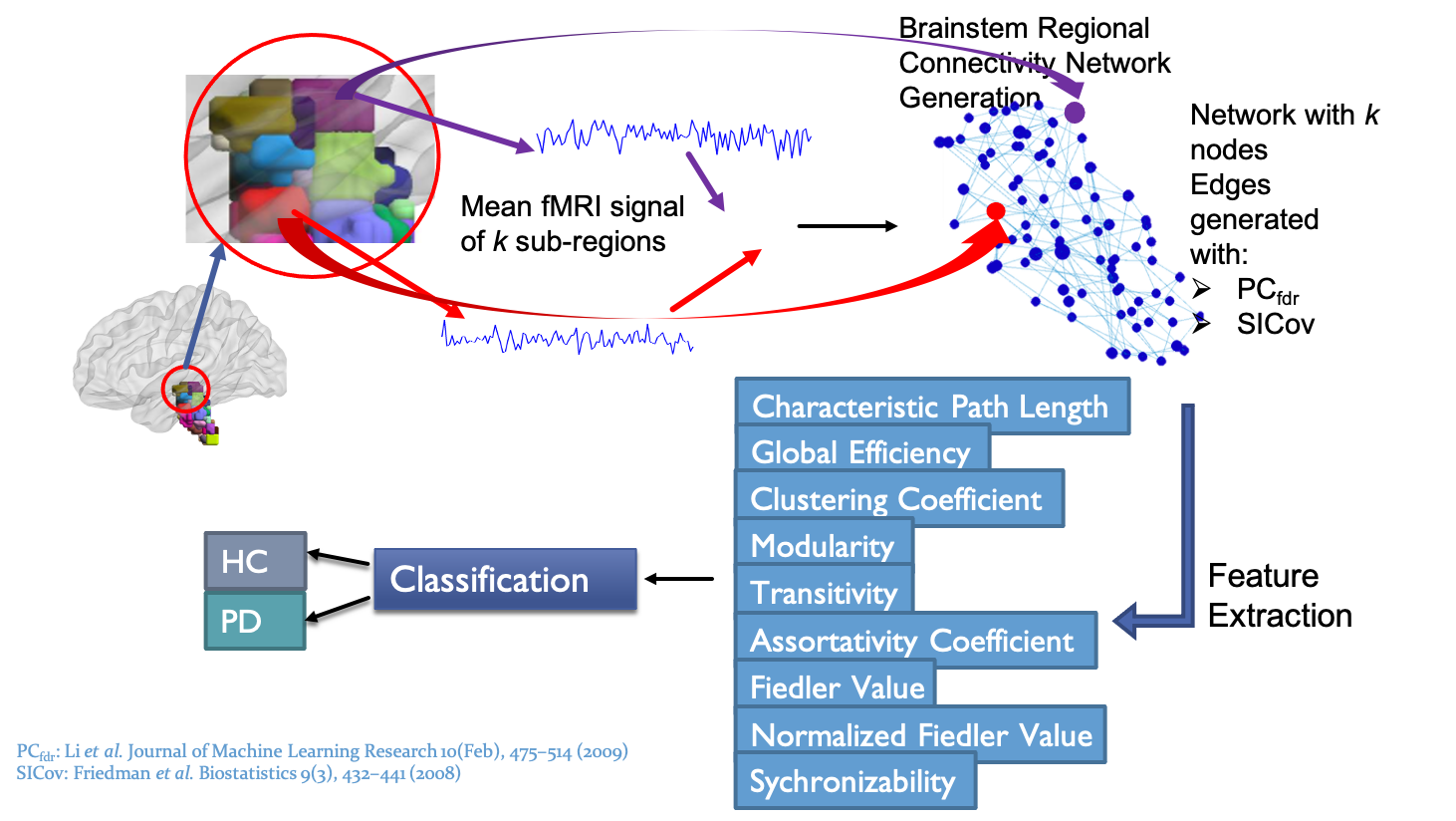
Parkinson's disease detection from brainstem
Prostate cancer detection from MRI
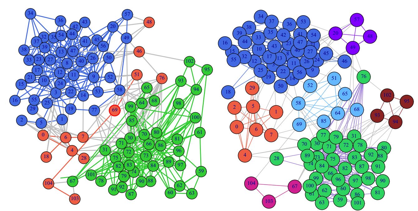
Unsupervised clustering from networks
Brain sub-region parcellation
Prostate cancer is the second most frequently diagnosed cancer and sixth leading cause of cancer-related death among males worldwide. Prostate cancer, if detected at an early stage, can be cured. Therefore an early stage diagnosis can play an important role in selecting the optimal course of treatment and hence reduce the mortality rate.
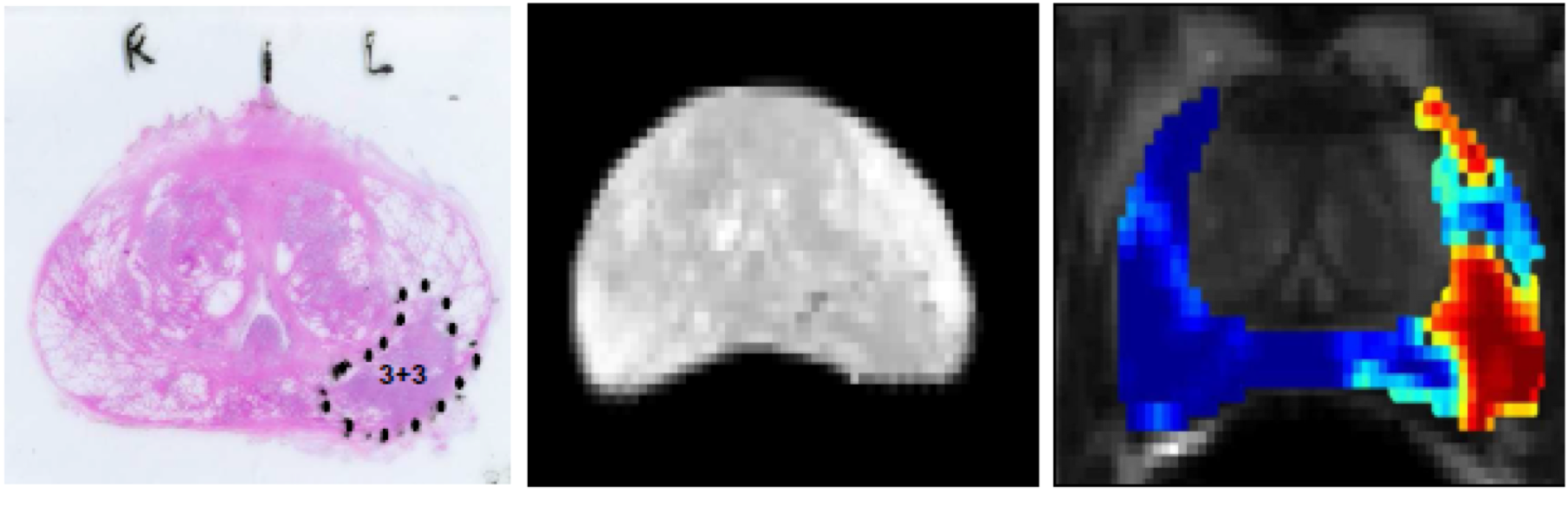
We proposed a novel set of data-driven features and a machine-learning framework to detect prostate cancer from Dynamic Contrast Enhanced (DCE) MRI images. The features are generated by mapping the DCE T1-weighted signal intensity to its principal component space. The optimal set of components is extracted with sparse regularized regression through a Least Absolute Shrinkage and Selection Operator (LASSO) model. A soft-margin support vector machine classifier was trained using the proposed features to generate cancer likelihood maps for the prostate gland. These likelihood maps show the likelihood of cancer for each pixel and hence highlight the regions from where the biopsy samples should be taken. The generated cancer likelihood maps have the potential to be used as a reference in MRI-targeted biopsies to decrease the possibility of missing clinically significant and potentially aggressive tumors.
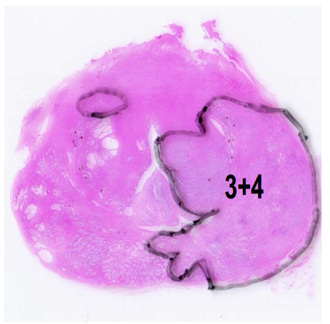
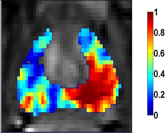
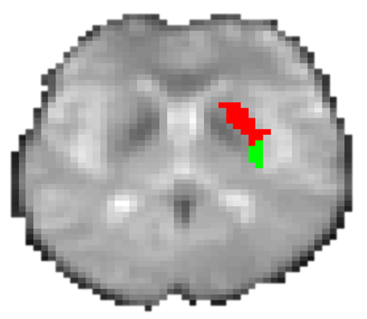
Left: Peripheral zone tumor marked in pathology slide (the ground truth). Right: The generated cancer likelihood map superimposed on the DCE T1-weighted image. The cancer likelihood map is generated from the DCE MRI signal.