|

|
|

Screening System for Deep Vein
Thrombosis (DVT)
|
|
|
|
With Ph.D. student Julian
Guerrero, co-supervised with Dr.
James McEwen, and clinical supervisors Drs. Bassam Masri and Dr. Savvas
Nicolau. Funded by NSERC and Delfi Medical.
 Phantom
video here.
Phantom
video here.
Carotid
segmentation video here.
|
DVT is a condition in which
blood clots forms in the veins of the legs. If dislodged, these blood clots,
called thrombi, can block the pulmonary circulation and cause death. DVT is
being diagnosed with ultrasound imaging, in an examination called compression
ultrasound (CUS). In this test, the veins are imaged with ultrasound. When
the operator of the ultrasound machine presses on the transducer, the vein
collapses under pressure, unless there is a thrombus inside. CUS consists
of repeated imaging compression cycles along the veins in search of thrombi.
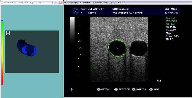
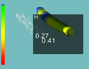
An objective, measurement-based approach to diagnosing DVT is being developed.
The ultrasound images are processed in order to locate, track and estimate
the cross-sectional venous area in real-time. At the same time, applied forces
by the ultrasound transducer are measured and the location of the utlrasound
transducer is obtained with an electromagnetic sensor. From the area and force
data, an objective estimate of vein complience can be obtained. From the
ultrasound transducer location, a map of the scanned veins can be obtained.
The result is segmented map of the veins that can be colour or otherwise code
to indicate venous compressibility and thus help the screeing process. The
figures below show the B-mode ultrasound image of a vessel phantom, is segmented
outline, the anatomical model built on segmented cross-sections and the overlayed
compressibility.
|
|
|
|
|
© 2001-2006 Tim Salcudean - last updated
November 2006
|
|