Computer Assisted Surgery with 3D Ultrasound
This is also a part of my PhD work which is about the design, realization, and evaluation of a three dimensional (3D) ultrasound (US) based minimally invasive computer assisted orthopedic surgery (CAOS) system for bone fracture assessment in emergency departments and bone fracture reduction assessment during orthopedic surgery. Our main goal is to create and develop a safe, acc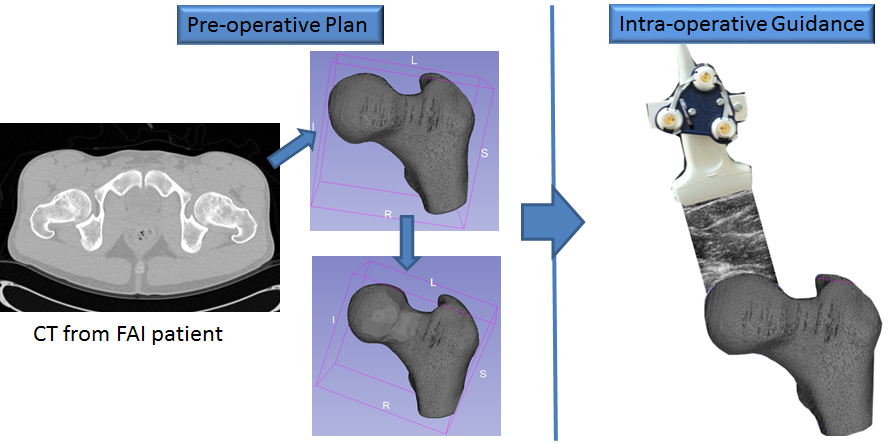 urate, robust and efficient system in which 3D US will be employed as and imaging modality to improve current state of the art CAOS systems. Such a system will ultimately address a variety of problems with the planning and execution of orthopaedic surgery procedures.
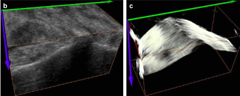
Ultrasound Image Segmentation and Feature Enhancement
Ultrasound (US) imaging has been extensively used in the medical field due to its non-invasive and non-destructive nature. It is the only portable intra-operative imaging modality with real-time, 3D, non-radiation based imaging capability. However US imaging has several limitations: user dependent image acquisition, limited field of view, imaging is orientation dependent, isoechoic properties of lesions relative to the adjacent soft tissues, the quality of the imaging is affected by the physical characteristics of the patient and overlying structures such as ribs, subcutaneous fat, and normal gas-containing structures. In order to use US in image guided interventions as an intra-operative imaging modality the relevant structures (bone, liver, prostate, and kidney) must be segmented with sufficient accuracy and speed. For this project I am interested in developing fast, accurate and automatic segmentation methods for extracting bone surfaces for orthoapedic applications and prostate boundary for prostate brachytherapy.
Automatic Image Registration for Image Guided Surgery
If images are to be useful in planning or guiding surgical procedures, they must be registered to the patient, such that image-space (defined by the 3-D array of voxels) can be related directly to the real-world coordinate system of the patient. This process is known as image registration. The ability to perform this registration accurately, automatically, and rapidly is critical for enabling more effective image guidance. Various techniques using these types of multimodal image fusion have been explored and have been shown to work with reasonable success. Linear registration techniques work well with sta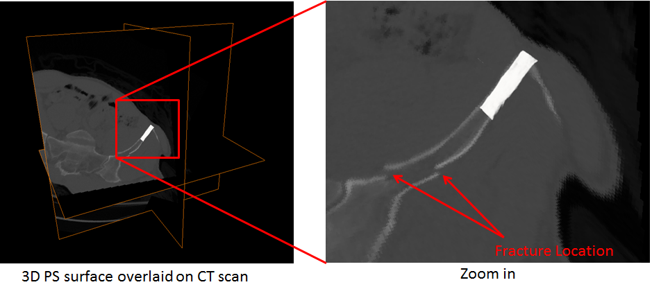 tic data but suffer when capturing anatomical movement. Nonlinear registration methods use complex mathematical transforms for data characterization and can be computationally intensive. For improving the quality of the fused image and 3-D visualization thereafter, other methods such as image segmentation, finite element modeling, or Bayesian algorithms need to be applied. Using sensors and markers to define fixed positions on the anatomy during image capture can also help with accurate alignment of pre- and intraoperative data. For this project, I am specifically interested in developing image registration methods in the frequency domain rather then in the intensity domain.