 Real-time
strain imaging of elastography phantom shown here.
Real-time
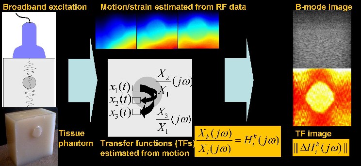
strain imaging of elastography phantom shown here.Vibro-elastography
is an extension of static elastography and was developed with Emre
Turgay and Rob Rohling. As illustrated below, the method consists
of applying broad-band low-frequency (typically <30Hz) compression
waves to tissue by a vibrator, measuring tissue displacement, and
either fitting a parametric model to the relative tissue motion, or
computing a frequency response from the relative tissue motion.
From
left to right, the figures below show: the low-frequency asymptotes of
these frequency responses in a soft-hard-soft cubic 5cm3
phantom (256x256), the conventional B-mode ultrasound image which does
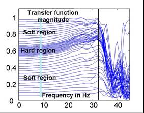
not show any contrast, and the magnitudes of the relative motion
responses of tissue at different depths relative to a tissue patch near
the vibrator (located at the top of
the images), as a function of frequency. Note
that in the hard region responses are closer together
and change more with frequency. With an 8MHz
transducer, stiffness changes of 10% and hard (twice
as stiff) inclusions of 0.5mm can be detected [Turgay,
Salcudean and Rohling 2006])
 |
 |
 |
See papers with Eskandari, Zahiri, Baghani for
recent work.