3D Ultrasound Resliced
The reslice plane including the epidural space, laminae and the color-coded line.
Abstract
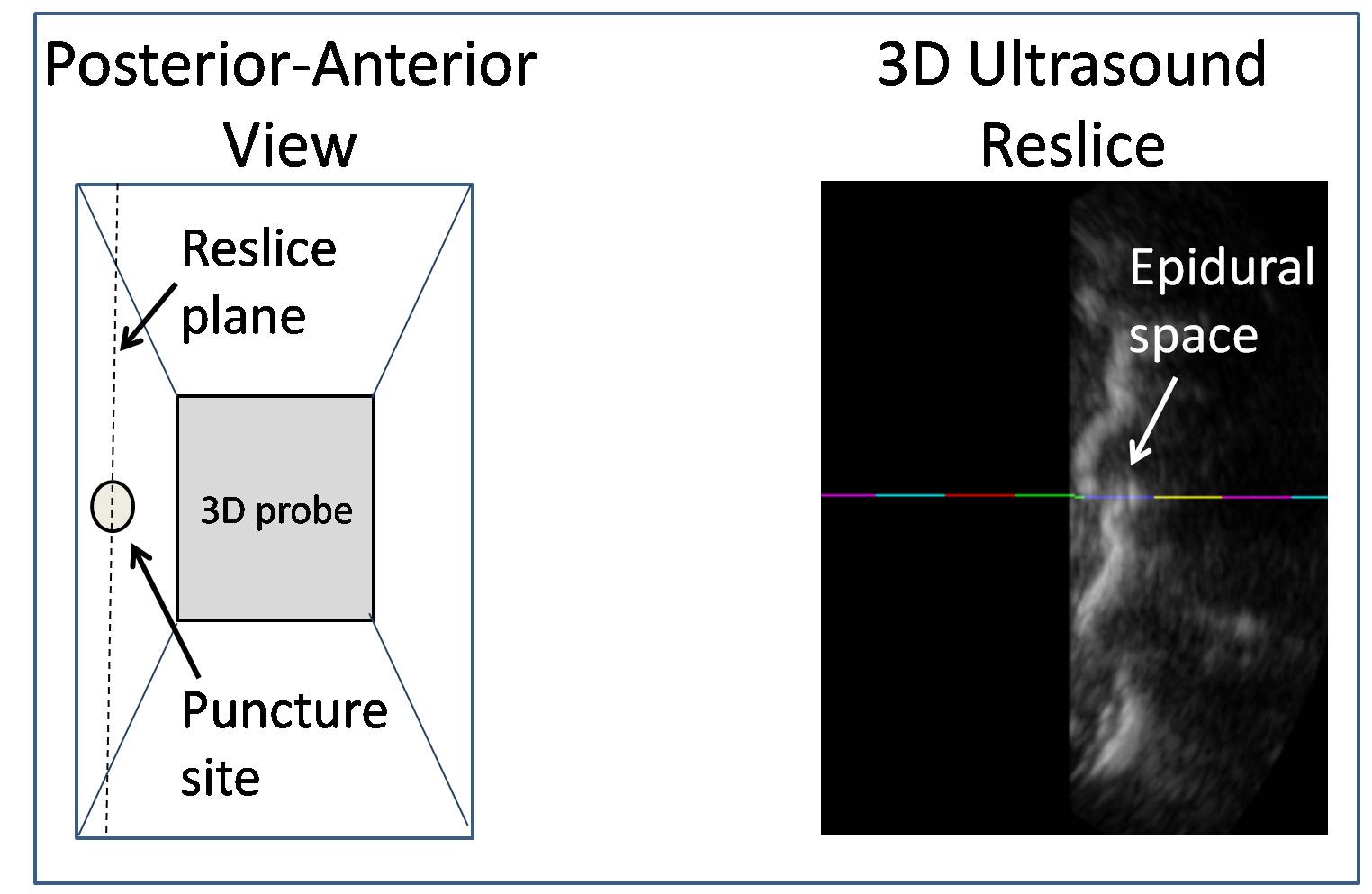
2D Ultrasound has previously been proposed as a pre-insertion imaging technique to measure the epidural depth from the skin surface as an aid for needle insertion. A new ultrasound technique is introduced for this task. The key innovation is the use of resliced image of 3D ultrasound to depict both the target anatomy and the anticipated midline needle path. Methods:ĀSlaughtered porcine subjects are obtained with the university animal care consent. The custom-designed approach includes a 3D ultrasound probe and a needle guide placed on the probe. A paramedian sagittal plane is extracted from the acquired 3D volume and displayed (Figure 3). This reslice plane is a virtual anterior-posterior view containing the needle path and the target epidural space. The target and path can be shown in ultrasound without having the transducer head obstruct the puncture site. The transducer is calibrated in a way that the anticipated needle path is depicted by a color-coded centimetric line on 3D resliced images.ĀIn experiments, the epidural space is found using the 3D resliced image. The epidural depth is measured using this image. The epidural depth is also measured using a standard 2D paramedian ultrasound technique. The epidural needle is inserted with the guide while the 3D resliced image is updated as each new 3D volume is acquired. Insertion is stopped upon loss-of-resistance to saline. The actual depth of needle insertion is also measured. Results:ĀInitial experiments were carried out on two recently slaughtered porcine subjects. The needle was inserted into all possible thoracic spaces (n=11 measurements). Comparing the 3D ultrasound reslice-measured depth and the actual needle depth, there is a bias of 5.4 mm and 95% limits of agreement of Bland-Altman -10.6mm to -0.2mm. Comparing the 2D paramedian ultrasound depth and the actual needle depth, there is a bias of 6.2 mm and 95% limits of agreement of Bland-Altman -11.7mm to -0.7mm. Discussion:ĀThe 3D ultrasound reslice gives comparable measurements to the traditional 2D paramedian ultrasound. The depth to the epidural space measured to the top of the echo from the ligamentum flavum is approximately 5mm, the thickness of the ligamentum flavum is a possible explanation for the bias. By adding the thickness of the ligamentum flavum, the limits of agreement for 3D reslice measurements becomes -5.6mm to 4.8mm.ĀIn summary, the 3D approach provides an acceptable framework to show ultrasound images of the thoracic epidural space and needle path together. Work is also progressing on providing a clear depiction of the needle in the 3D resliced image as it is inserted.Papers
Porcine thoracic epidural depth measurement using 3D ultrasound resliced images.
Canadian Anesthesiologists Society Annual Meeting,
2011.
Dual-source ultrasound identification of the lumbar epidural space allows real-time guidance of midline lumbar epidural needle insertion in a porcine model
Society for Obstetric Anesthesia and Perinatology (SOAP) 42nd Annual Meeting,
2010.
Abtin Rasoulian Last modified: Nov 5, 2012