Spine Segmentation from CT Images
Abstract
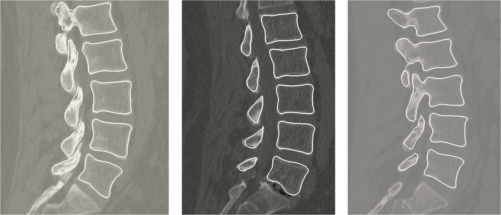
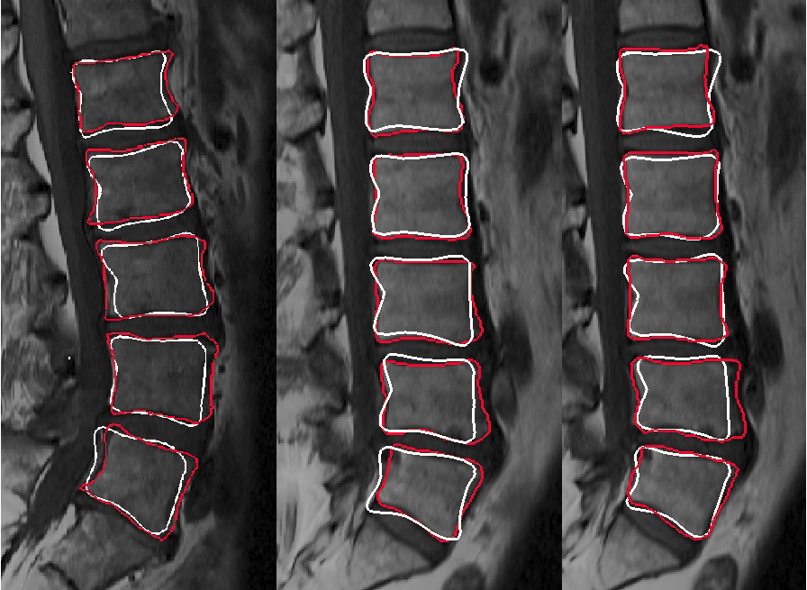
Segmentation of the spinal column from medical images such as CT and MR is a pre-processing step for a range of image guided interventions. Current techniques focus on identification and separate segmentation of each vertebra. Recently, statistical multi-object shape models have been introduced to extract common statistical characteristics between several anatomies. These models are shown to be robust and accurate for segmentation purposes. In this paper, we reconstruct a statistical multi-vertebrae shape+pose model and propose a novel technique to register such a model to CT images.
Papers
A statistical multi-vertebrae shape+pose model for segmentation of CT images
SPIE Medical Imaging,
2013.
Lumbar Spine Segmentation Using a Statistical Multi-vertebrae Anatomical Shape+Pose Model
IEEE Transactions on Medical Imaging,
2013.
Volumetric MR Images Using a Statistical Shape+Pose Model
SPIE Medical Imaging,
2014.
Abtin Rasoulian
Last modified: Nov 5, 2012